

Your Byron Eye Doctor
About EyeCare Dimensions
EyeCare Dimensions has been dedicated to serving the community of Byron since 1987, offering exceptional and individualized eyecare services for patients spanning all age groups. Our team at EyeCare Dimensions is committed to conducting thorough eye examinations, providing expert contact lens fittings, managing eye injuries, infections, and glaucoma, and screenings for cataracts and various other eye conditions.


Our Eye Care Services

Comprehensive Eye Exams
From updating your eyeglasses prescription to detection and treatment of eye diseases, comprehensive eye exams are important for continued visual health.

Contact Lens Exams
During a contact lens exam, your eye doctor will check if you are a good candidate for contacts and find the best type of contacts for your needs..

Emergency Eye Care
Eye emergencies such as severe pain in the eye, or foreign objects puncturing the eye require immediate attention. Contact us immediately.
Over 50% of people living with glaucoma don’t know they have the disease as it shows no symptoms. Early detection and treatment can prevent blindness.
Eyeglasses & Frames

Designer Frames
Our extensive optical section offers a wide variety of eyeglass frames in every style, material & design. Come visit us today to see for yourself!
Our expert optical team can find just the right pair of glasses for you to be confident and look your best.
Lens coatings improve visual comfort, make it easier to clean your glasses and ensure your lenses last longer. Coatings include anti-scratch, anti-reflective, photochromatic and UV / blue light filters.
Contact Lenses

Contact Lens Fitting
We offer a wide range of contact lens options from dailies, monthly to multifocal contact lenses for crystal clear vision and superior comfort.
During a contact lens exam, your eye doctor will check if you are a good candidate for contacts and find the best type of contacts for your needs.
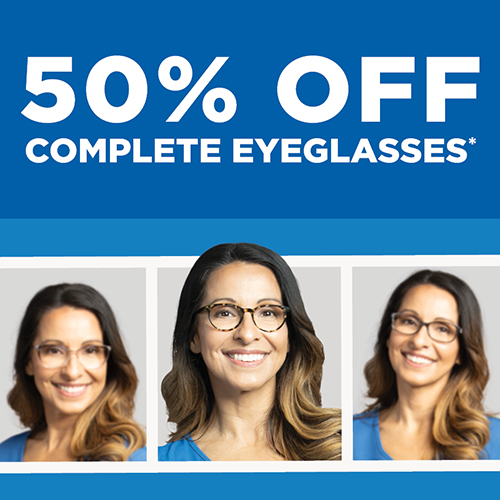
Buy One, Get One 50% Off Eyeglasses.
*Requires purchase of complete prescription pairs, including frame and lenses. Discount applied to complete pair of equal or lesser value. Does not include sunglass frames,Barton Perreira, Cartier, Cazal, Chanel, Cutler and Gross, Dior, Dita Lancier, Fendi, Gucci, ic!Berlin, l.a. Eyeworks, Maui Jim, Mykita, Nifties, Oakley, Oliver Peoples, Persol, Ray-Ban, Robert Marc, Salt, Salvatore Ferragamo, Skaga, Silhouette, Tom Ford, WOOW, accessories, contact lenses, or medical procedures. Cannot be combined with any other discounts, promotions, or insurance plans. Not valid on previous orders. Other restrictions may apply. See practice for full details. Offer valid 04/08/2024-06/16/2024. 24AEG-729313


Location & Opening Hours
On the corner of Blackhawk Drive and Chestnut Street. Across the street from Costa's Pizzeria & Ristorante.
- Monday 9:00 am - 5:00 pm
- Tuesday 9:00 am - 6:00 pm
- Wednesday Closed
- Thursday 9:00 am - 6:00 pm
- Friday 9:00 am - 5:00 pm
- Saturday 9:00 am - 1:00 pm
- Sunday Closed